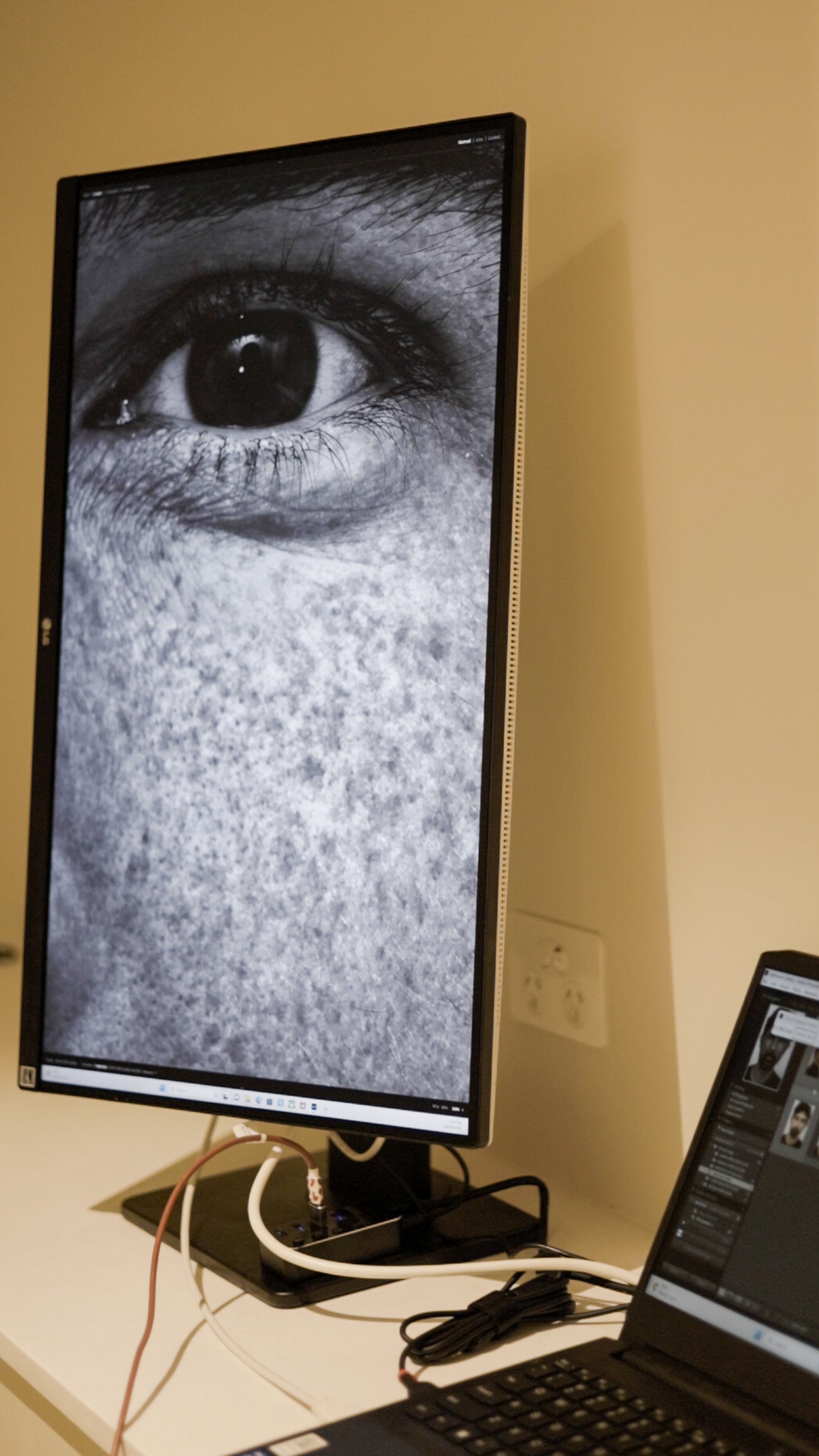
Skin Filters
In 2014, I first encountered a UV woods lamp and noted its potential for replication through the utilization of raw photography data acquired from a calibrated camera under specific conditions.
In 2017, we established computer code to extract this acquired data, incorporating an analysis of both photo damage and vascularity. Over time, this development has proven to be an invaluable tool for dermatologists, dermal therapists, and injectors in their examination of patient skin.
Although "skin health" is not a concept that immediately springs to mind for most patients, it is, in fact, the fundamental purpose for capturing these images – to document the skin's overall health.
This documentation includes aspects such as its luminosity, plumpness, and degree of wrinkling, which can all be effectively showcased using these filters.
Our Melanin filter is specifically designed to display the concentration of melanin within the epidermis, while our vascular filter highlights areas where blood vessels are located closest to the skin's surface.
Conditions that can be showcased :
Solar keratosis
Photoaging
Hyper pigmentation
Rosacea
Melasma
Dermatitis
Acne
Vitiligo
Ketatosis Pilaris ( Woodrow has this not fun! )
Eczema
Hypertrophic scarring
Rhinophyma
Spider Veins
Varicose veins


